
Valeriia Kolotusha
Ukraine
Approach to P16 and Ki-67 in the Cervical Intraepithelial Neoplasia staging
Valeriia Kolotusha1, Viktoriia Khoperiia1
1. Taras Schevchenko National University of Kyiv
Abstract
Background
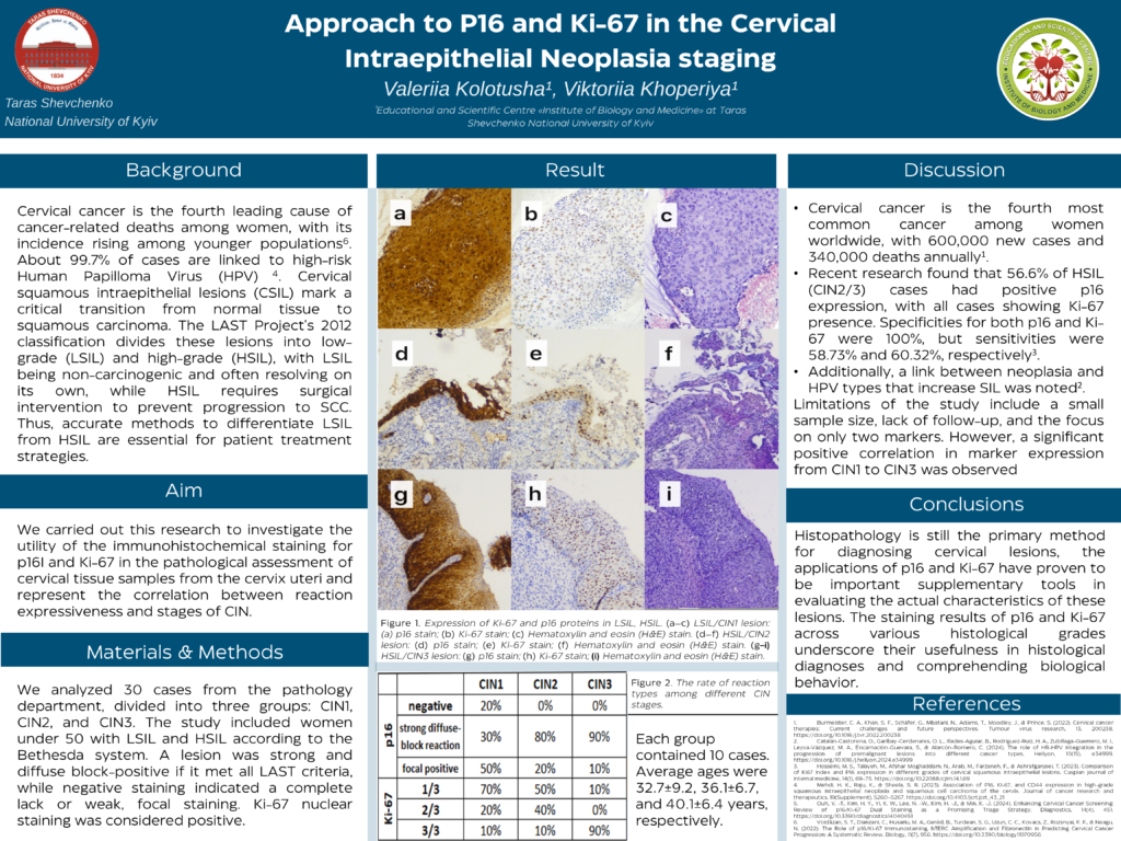
Cervical cancer is the second most common malignancy in women, which originates from cervical intraepithelial neoplasia (CIN). Accurate screening and diagnosing are increasingly in demand for early detection and prevention of precancerous lesions. Human papillomavirus (HPV) infection has been identified as the leading cause of cervical neoplasia. CIN is categorized into CIN1, CIN2, and CIN3 depending on the degree of epithelial involvement. To improve differentiation between CIN 1 and CIN2/3 IHC markers P16 and Ki-67 are used. Hyperproduction of P16 is associated with HPV, but some normal cervical tissues express P16. The Ki-67 antibody detects proteins that are expressed in cell proliferation. Thus, Ki-67 expression and P16 staining are recommended for identifying high-risk precursor lesions and cervical cancers. This study aimed to evaluate the significance of p16 and Ki67 immunohistochemical staining in the assessment of cervical squamous intraepithelial lesions.
Methods
Women who underwent biopsy were retrospectively included in the study. All histological samples were processed according to routine procedures. H&E-stained and IHC slides of all biopsy samples were reviewed and classified according to the criteria outlined by the LAST project.
Results
We found, that p16 expression is divided into three groups: negative, heterogenic positive and diffuse nuclear-cytoplasmic positive reaction. In cases that are histologically classified as CIN1, p16 showed inconsistent positivity with a moderate intensity. The glandular epithelium displayed clear signs of atypia, and p16 revealed inconsistent moderate positivity. In all CIN stages, Ki-67 was nuclear positive in the basal and parabasal layers and different parts of the squamous epithelium. The results showed significant differences in p16 and Ki67 expression among normal and CIN. The expression levels of P16 and Ki-67 showed a positive correlation with the severity of cervical lesions.
Conclusions
Consequently, utilizing both P16 and Ki-67 can help identify patients at a higher risk for SCC and potentially decrease misdiagnosis rates, making it highly valuable for differentiating between SCC and CSIL in younger women. However, further research with a larger sample size is required to validate this conclusion.
Leave A Comment